Dr V. W. Verlekar

What Is MRV?
NEUROLOGY
6/20/20256 min read

Introduction to MR Venography
Magnetic Resonance Venography (MRV) is a specialized imaging technique employed to evaluate the venous drainage abnormalities of the brain. MRV provides a comprehensive view of the cerebral venous system without the need for invasive procedures, offering a safe and efficient method for diagnosis.
Techniques Used in MR Venography
Two primary techniques are utilized in MR venography: 2-dimensional (2D) time-of-flight (TOF) and 3-dimensional (3D) phase-contrast (PC) imaging. Both methods are preferred due to their ease of use and the lack of requirement for contrast agents.
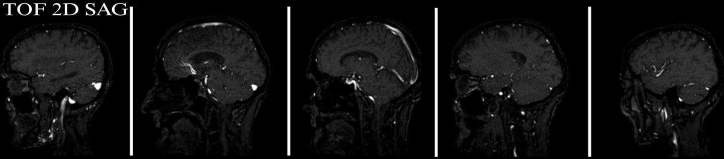
The 2D time-of-flight technique is particularly effective for visualizing the venous sinus structures by using a series of rapid imaging sequences. It captures clear images of the blood flow in the veins, making it easier to identify any abnormalities.
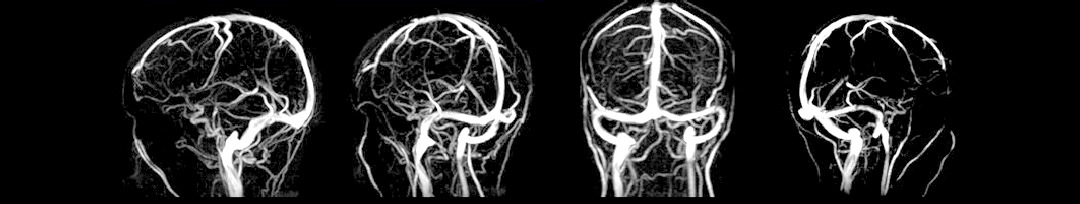
On the other hand, the 3D phase-contrast method offers a volumetric acquisition of images, providing a more detailed assessment of the cerebral venous sinuses. This technique is valuable in quantifying blood flow and delineating small structures within the venous system, enhancing diagnostic accuracy.
TI ME-OF-FLIGHT (TOF)
These techniques show contrast between stationary tissues and flowing blood by manipulating the magnitude of magnetization.
The magnitude of magnetization from the moving spins is very large compared to the magnetization from the stationary spins which is relatively small. This leads to a large signal from moving blood spins and a diminished signal from stationary tissue spins. Time of flight (TOF) utilizes the longitudinal magnetization vector for imaging.
PHASE CONTRAST (PC)
Phase contrast scans can be used for 2D or 3D imaging.
These techniques derive contrast between stationary tissues and flowing blood by manipulating the phase of the magnetization. The phase of the magnetization from the stationary spins is zero and the phase of the magnetization from the moving spins is non- zero. The phase is a measure of how far the magnetization process from the time it is tipped into the transverse plane until the time it is detected. A bipolar gradient pulse with equal magnitude and opposite direction is used to diminish signal from the stationary tissue. Phase contrast angiography (PCA) utilises the transverse magnetization vector. In phase difference images, the signal is linearly proportional to the velocity of the spins. Fast moving spins give rise to a larger signal and spins moving in one direction are assigned a bright signal and appear white in the scan whereas spins moving in the opposite direction are assigned a dark signal and appear black in the scan. Phase contrast methods are sensitive to a range of velocities, so the user must choose this value carefully. Different velocity encoding values can be used in different scans to highlight different vessels. High velocity encoding for arteries (40-70 cm/sec) due to arterial flow is fast. Low velocity encoding for veins (10-20 cm/sec) due to venous flow is slow.
INDICATIONS FOR MAGNETIC RESONANCE VENOGRAPHY (MRV) BRAIN
1. Evaluation of thrombosis
2. Tumour of the cerebral venous sinus
3. Drowsiness and confusion accompanying a headache
CONTRAINDICATIONS MAGNETIC RESONANCE VENOGRAPHY (MRV) BRAIN
1. Any electrically, magnetically or mechanically activated implant (e.g. cardiac pacemaker, insulin pump biostimulator, neurostimulator, cochlear implant, and hearing aids).
2. Intracranial aneurysm clips (unless made of titanium)
3. Pregnancy (risk vs benefit ratio to be assessed)
4. Ferromagnetic surgical clips or staples
5. Metallic foreign body in the eye
6. Metal shrapnel or bullet
PATIENT PREPARATION MAGNETIC RESONANCE VENOGRAPHY (MRV) BRAIN
1. A satisfactory written consent form must be taken from the patient before entering the scanner room.
2. Ask the patient to remove all metal objects including keys, coins, wallet, cards with magnetic strips, jewellery, hearing aid and hairpins.
3. If possible provide a chaperone for claustrophobic patients (e.g. relative or staff).
4. Offer earplugs or headphones, possibly with music for extra comfort.
5. Explain the procedure to the patient
6. Instruct the patient to keep still
7. Note the weight of the patient
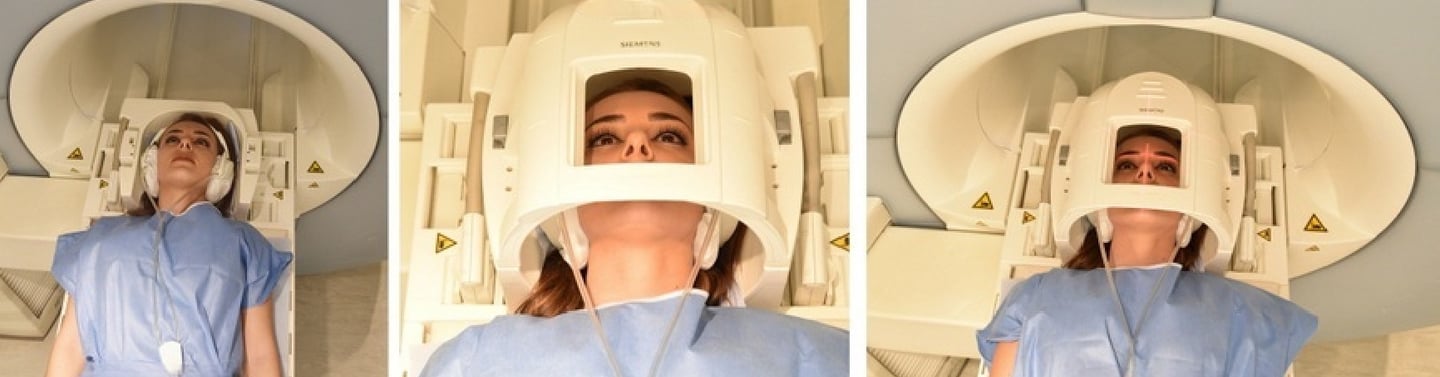
POSITIONING OF PATIENT FOR MAGNETIC RESONANCE VENOGRAPHY (MRV) BRAIN
1. Head first supine.
2. Position the head in the head coil and immobilise with cushions.
3. Give cushions under the legs for extra comfort.
4. Centre the laser beam localiser over the glabella
Suggested Protocols, Parameters, and Planning for MRI Scanning
#### Localiser
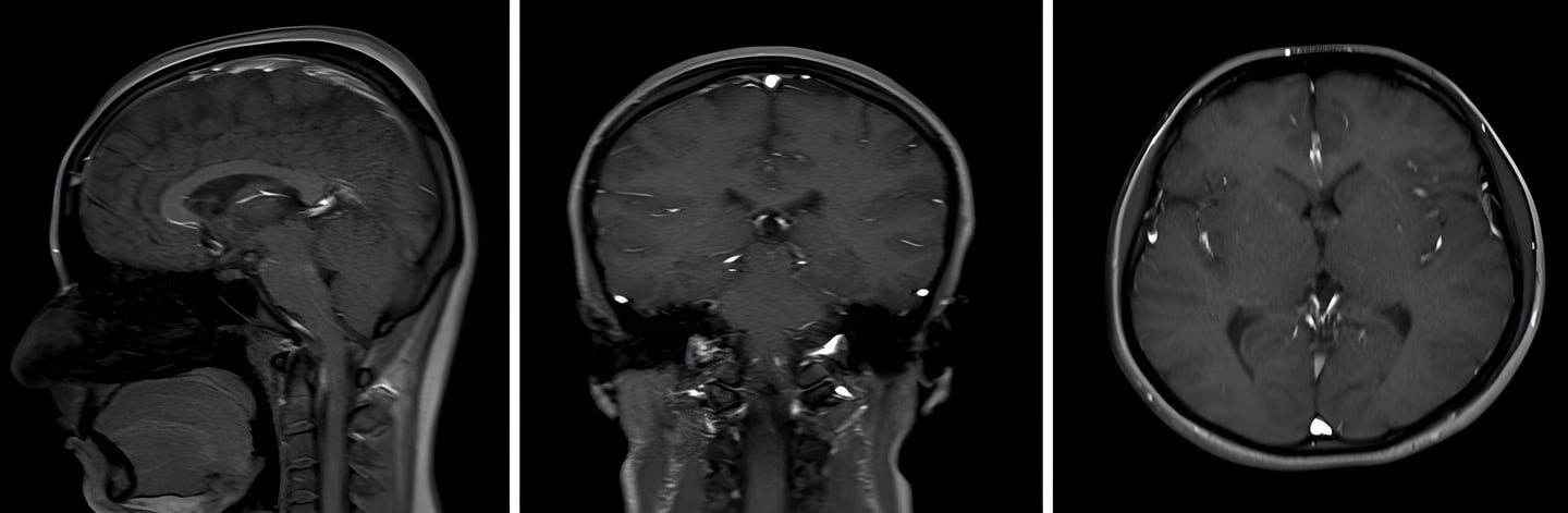
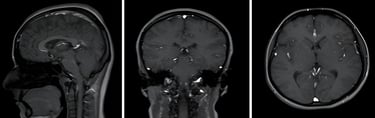
Sequence To initiate the MRI scanning process, a three-plane localizer should be conducted. This localizer is critical for accurate localization and planning of subsequent sequences. The localizer scans are typically T1-weighted, low-resolution scans that require less than 25 seconds to complete. This time efficiency allows for rapid assessment and ensures that patient positioning is optimized before more detailed imaging is performed.
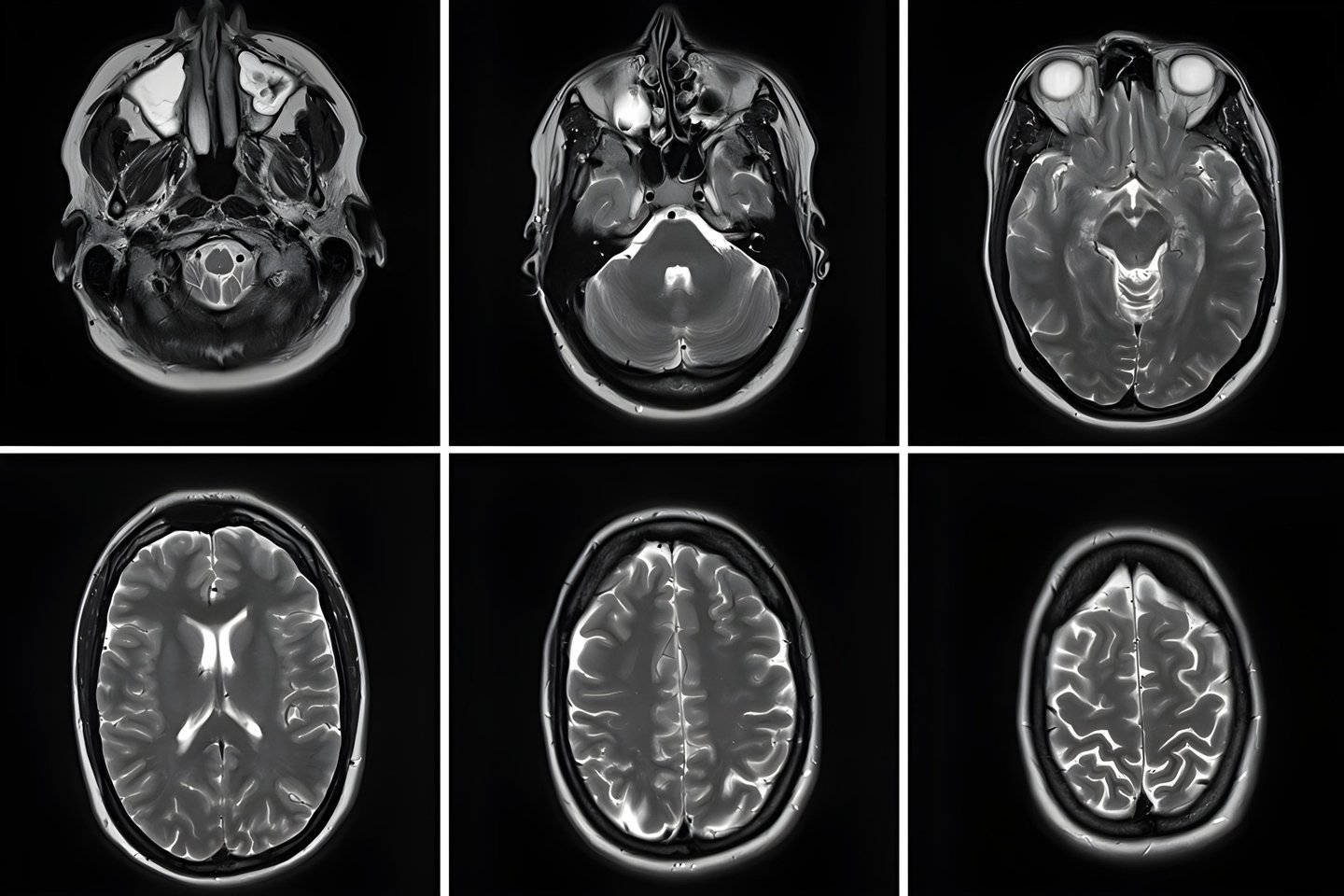
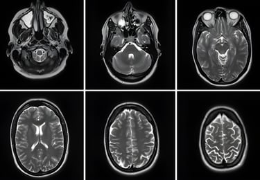
#### T2 TSE Axial Imaging
Following the localizer sequences, the next step involves the acquisition of T2 turbo spin echo (TSE) axial images. The planning of these axial slices should be carefully executed on the sagittal plane. It is essential to angle the positioning block parallel to the genu and splenium of the corpus callosum to ensure comprehensive brain coverage.
1. Slice Coverage: The slices must encompass the entire brain, extending from the vertex to the line of the foramen magnum. This ensures that no critical areas are omitted from the imaging.
2. Positioning Check: After setting up the axial slices, it is imperative to verify the positioning block in the other two planes (coronal and axial). This step is vital for confirming that the imaging planes are correctly aligned and optimizing the quality of the images to be obtained.
3. Angle Adjustment: Care must be taken to provide an appropriate angle in the coronal plane, particularly when dealing with a tilted head. The block should be set perpendicular to the line of the third ventricle and the brain stem. This positioning is crucial for achieving accurate anatomical representation and diagnostic quality in the final images.
In summary, the meticulous planning of MRI sequences using localizers and detailed axial imaging strategies is essential for obtaining high-quality diagnostic images, facilitating better clinical assessments.
2D TOF or 3D PC
The 2D Time of Flight technique is a widely utilized method in MRV due to its ability to visualize blood flow without the need for contrast agents. This approach leverages the inherent properties of magnetic resonance imaging (MRI) to capitalize on the differences in signal intensity between static tissues and flowing blood. In 2D TOF MRV, images are acquired in a series of slices perpendicular to the direction of blood flow. The primary advantage of this technique is its high spatial resolution and the capacity to produce detailed images of the venous anatomy. The sequences used typically involve a short repetition time (TR) and a short echo time (TE), optimizing the visualization of vessels, particularly in regions with complex anatomy, such as the brain or neck. However, this technique is not without its limitations. It can suffer from susceptibility artifacts, particularly in areas with high magnetic field strengths or near air-tissue interfaces, which can obscure vessel borders. Additionally, 2D TOF MRV can be less effective for deep venous structures where the flow may be slow or complex.
3D Phase Contrast (PC) MRV On the other hand offers a different approach, focusing on measuring the velocity of blood flow. This method enables the visualization of both the morphology of vessels and the dynamics of blood movement within them. 3D PC MRV is particularly beneficial in scenarios where understanding the flow dynamics is crucial, such as in assessing vascular malformations or planning surgical interventions. In 3D PC MRV, a series of images is collected in a volumetric manner, allowing for reconstruction in multiple planes. Unlike TOF, the phase contrast technique relies on the magnetic properties of flowing blood, providing both qualitative and quantitative data about flow rates. This feature is particularly useful in cases where there's a need to evaluate conditions such as venous stenosis or thrombosis. The challenge with 3D PC MRV lies in the complexity of interpretations, as the acquisition and analysis of flow data require a sound understanding of hemodynamics. Additionally, the technique can be susceptible to various artifacts, including aliasing, which may distort the flow measurements.
Comparative Analysis
When choosing between 2D TOF MRV and 3D PC MRV, it is essential to consider the clinical context and the specific information required. 2D TOF is often favored for straightforward anatomical assessments and is generally quicker to perform, making it suitable for emergency scenarios. Conversely, 3D PC MRV shines in situations that necessitate detailed flow characterization, providing both anatomical and hemodynamic insights. In practice, these two techniques can complement each other. A two-step approach where a quick 2D TOF assessment is followed by a more detailed 3D PC evaluation may yield a comprehensive understanding of vascular conditions, benefiting both diagnostic accuracy and patient management. In summary, the choice between 2D Time of Flight and 3D Phase Contrast MRV comes down to the specific clinical requirements, the anatomical region of interest, and the nature of the suspected vascular pathology.